Mantis Micrographs

Mantidforum

Help Support Mantidforum:

This site may earn a commission from merchant affiliate links, including eBay, Amazon, and others.

jfmantis

Well-known member
Joined
May 2, 2007
Messages
58
Reaction score
0
Location
NY
I have decided to post my collection of mantis micrographs. A micrograph is a picture taken by a microscope. A few of them have poor resolution, but it was the best I could do. Enjoy!

First up, the pictures of dead nymphs. They are either Stagmomantis californica, or Iris oratoria, I don't know which. These are all L1 and from the same ootheca.

Nymphside60x.jpg


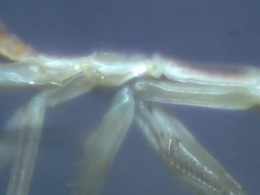
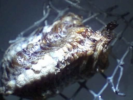
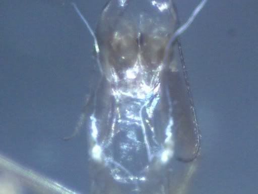
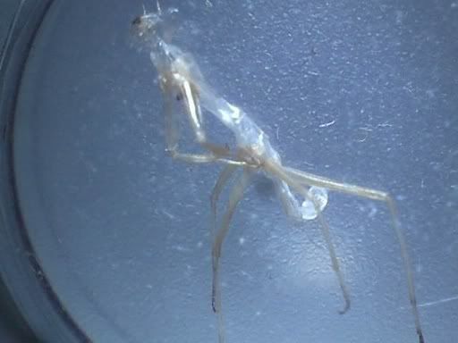
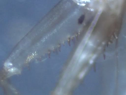
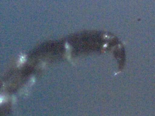
Side of a nymph at 60x. You can see the forelegs.

Nymphhead60x.jpg


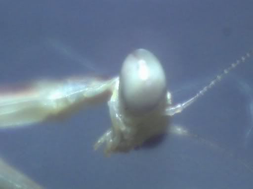
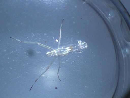
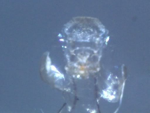
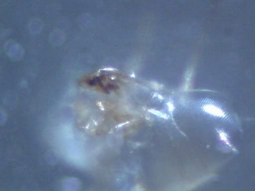
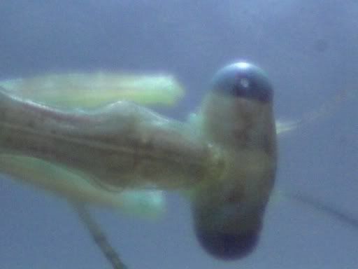
Head of a nymph at 60x. It sort of seems like it is looking at you.

Nymphbehind200x.jpg


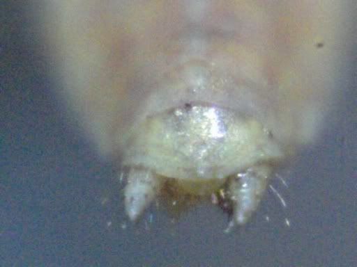
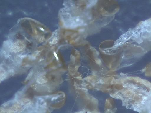
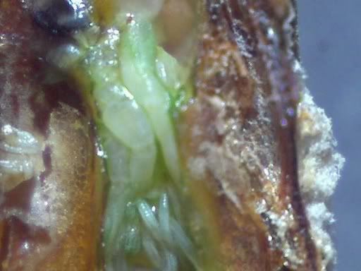
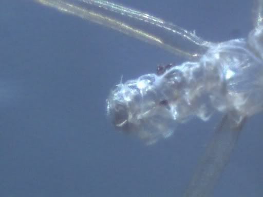
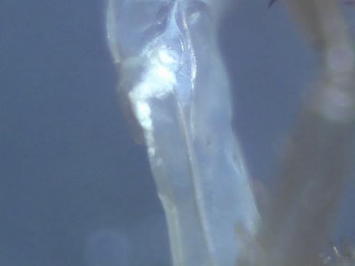
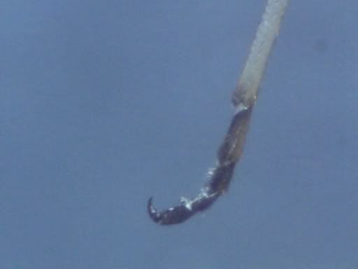
"Posterior" of a nymph at 200x.

Nymphantennae60x.jpg


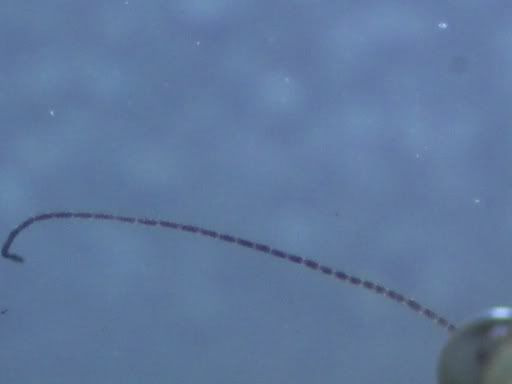
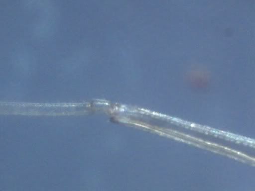
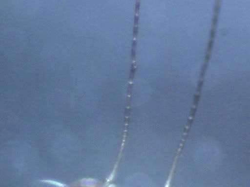
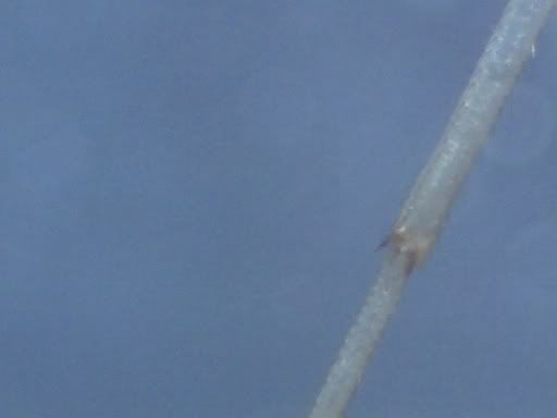
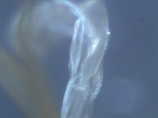
Antennae, at 60x. I wonder how many segments they have?

Nympheye200x.jpg


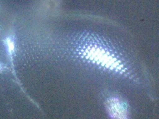
The eye, at 200x. Can you see the lenses in the eye?

Well, thats all for those unfortunate little guys. :(

More pictures to come!

 
Now, for my ootheca pictures:

Mantis_ootheca__10x.jpg


Ootheca at 10x. This one was from a wild caught mantis. It only hatched 3 nymphs. It is either Iris oratoria or Stagmomantis californica. Does anybody know?

Tasoothecadust60x.jpg


I don't really know what to call this, but it is at 60x. When my Chinese mantis

oothecae hatched, this was the powdery sawdust like stuff that hung out.

Insideofootheca60x.jpg


Inside of ootheca at 60x. When I cut open an ootheca (picture #1) to see if it would hatch, this is what I saw. You can see the nymph shape. It was also pulsating.

 
Oops. Right after I posted the ootheca pictures, I realized that I had forgotten to add my other pictures too. :oops: Here they are:

My large collection of pictures of molts:

The following are all pictures of 1 molt. It is L1 to L2 exoskeleton. Again, either S. californica or I. oratoria.

Wholebody10x-1.jpg


The whole molt at 10x

Neck60x-1.jpg


This is the 'neck' (thorax?) at 60x.

Leg60x.jpg


First joint down on the leg at 60x. Hairy legs :) .

Head60x-1.jpg


Head, at 60x.

Behind60x.jpg


Posterior, at 60x. The abdomen always is very thin in molts.

The next molts are from (of course), either S. californica, or I. oratoria. This is the L3 to L4 exoskeleton.

Wholebody10x.jpg


Whole body at 10x.

Antennae60x.jpg


Antennae, at 60x.

Neck60x.jpg


'Neck' at 60x. The dark blur on the right is one of the forelegs.

Legjoint160x.jpg


First down leg joint at 60x.

Legjoint260x.jpg


Second down leg joint at 60x. It looks like the joint has spines on it.

Head60x.jpg


Head, at 60x. The mouth has dried bug blood on it.

Forelegs60x.jpg


Forelegs at 60x. It is amazing how detailed the molts are!

Eye200x.jpg


Eye at 200x. Here you can really see the lenses in the compound.

Clawclose-up200x.jpg


Claw at end of leg, at 200x. This one is about 150 microns long.

Clawatendofleg60x.jpg


Claw at end of leg, at 60x. You can see the whole tarsus now.

Abdomen60x.jpg


Abdomen, at 60x. So skinny.

That is all for my mantis micrographs, when I get more, I will post them :)

 
Thanks :) .

I finally determined the species of my mantids! They are all Stagmomantis californica (except for ootheca picture #2)...I think. :(

Also, I appreciate your compliments about my "lens" robomantis, but I'm not using a camera. I took them with a microscope.

Finally, here is a picture I forgot. It is one of the dead nymph pictures:

Backofhead60x.jpg


Back of head, at 60x.


Latest posts

Top